PrognoHealth – Corporate Health & Wellness Specialist

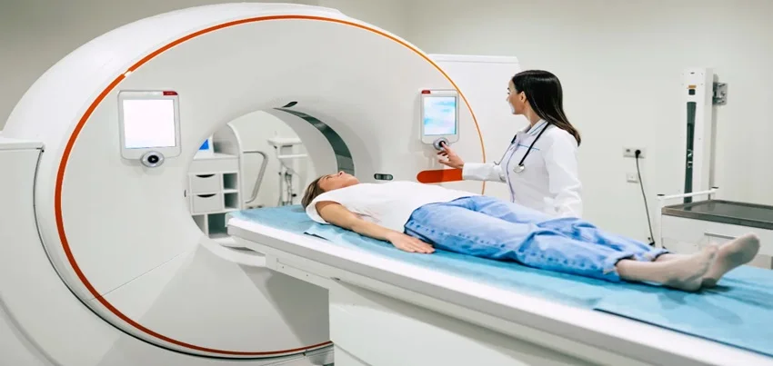
CT Scan of the Abdomen and Pelvis A CT scan of the abdomen and pelvis is a medical imaging test that combines X-ray technology with computer processing to create detailed images of the internal organs and tissues in these regions. This diagnostic test can help doctors detect a wide range of conditions, such as tumors, infections, inflammation, or injuries, that may affect the organs in the abdomen and pelvis, including the liver, kidneys, pancreas, spleen, bladder, and reproductive organs. In this blog post, we will discuss how this test is conducted, what are the common symptoms for ordering the test, how to prepare for the test, how long does it take, and how the results are interpreted. Test procedure and preparation : Before the CT scan, the patient may be asked to remove any metal objects, such as jewelry or dentures, and to wear a hospital gown. The patient will then lie down on a table that slides into a large, doughnut-shaped machine that emits X-rays. The machine will take a series of cross-sectional images of the abdomen and pelvis, which will be processed by a computer to create detailed, 3D images. During the scan, the patient may be asked to hold their breath or to stay still to avoid any blurring of the images. In some cases, the doctor may require the use of contrast dye, which helps highlight the organs and blood vessels in the abdomen and pelvis. The contrast dye may be given orally, intravenously, or through an enema. Before the test, the patient may be asked to fast for several hours, to avoid eating or drinking anything that may interfere with the absorption of the dye. Common symptoms for ordering the test : A CT scan of the abdomen and pelvis may be ordered by a doctor if the patient is experiencing symptoms such as abdominal pain, bloating, diarrhea, constipation, vomiting, blood in the urine or stool, or unexplained weight loss. This test may also be used to evaluate the effectiveness of a treatment for a known condition, to monitor the progression of a disease, or to screen for certain cancers, such as colon or ovarian cancer. Time taken for the test and results interpretation : The duration of the CT scan of the abdomen and pelvis depends on the complexity of the images required and whether or not a contrast dye is used. Typically, the test takes between 30 minutes to an hour to complete. After the test, the images will be reviewed by a radiologist, who will provide a report to the doctor. The doctor will then discuss the results with the patient, which may include further testing, treatment, or referral to a specialist. A CT scan of the abdomen and pelvis may be included in a comprehensive health checkup, which is a series of medical tests and screenings that are performed to evaluate a person’s overall health and well-being. A health checkup may also include blood tests, urine tests, electrocardiograms, and other imaging tests, such as a mammogram or a bone density scan. In the context of corporate health and wellness, a CT scan of the abdomen and pelvis may be offered as part of an employee health program, which aims to promote the health and productivity of the workforce. This program may include regular health screenings, wellness coaching, fitness classes, and other health-related services. In conclusion, a CT scan of the abdomen and pelvis is a valuable diagnostic tool that can help doctors detect and diagnose a wide range of conditions affecting the organs in these regions. If you are experiencing any symptoms that may indicate a problem in the abdomen or pelvis, or if you are due for a routine health checkup, speak with your doctor about whether a CT scan may be appropriate for you. “Comprehensive Guide to CT Scan of Abdomen and Pelvis: Procedure, Benefits, and Applications Introduction: A CT scan of the abdomen and pelvis is a valuable diagnostic tool used to evaluate various abdominal and pelvic conditions. This comprehensive guide provides insights into the importance, procedure, benefits, and applications of CT scans in assessing abdominal and pelvic health. Importance of CT Scan of Abdomen and Pelvis: CT scans of the abdomen and pelvis play a crucial role in diagnosing and evaluating a wide range of medical conditions affecting these regions. From detecting kidney stones and appendicitis to staging tumors and assessing traumatic injuries, CT scans provide detailed images that aid in accurate diagnosis and treatment planning. Procedure for CT Scan of Abdomen and Pelvis: Before the CT scan, patients may be required to fast for a few hours and refrain from drinking fluids to ensure optimal imaging quality. During the procedure, the patient lies on a table that moves through a doughnut-shaped scanner. Contrast material may be injected intravenously to enhance visualization of certain structures. Patients are advised to remain still during the scan to obtain clear images. Precautions and Preparation: Patients should inform their healthcare provider about any allergies, pre-existing medical conditions, or medications they are taking, especially if they have kidney problems or diabetes. It’s essential to follow all instructions provided by the healthcare team regarding fasting, fluid intake, and medication adjustments before the scan. Results Interpretation: After the CT scan, radiologists interpret the images to identify any abnormalities or signs of disease. The results are typically shared with the referring physician, who will discuss the findings with the patient and recommend further diagnostic tests or treatment options if necessary. CT Scan of Abdomen and Pelvis vs. MRI: While both CT scans and MRI provide detailed images of the abdomen and pelvis, they use different imaging techniques. CT scans use X-rays to create cross-sectional images, making them ideal for detecting bone abnormalities and evaluating solid organs. On the other hand, MRI uses magnetic fields and radio waves to produce images, offering superior soft tissue contrast and avoiding ionizing radiation. Benefits of CT Scan of Abdomen and Pelvis: Rapid and non-invasive imaging High-resolution images for accurate
Read More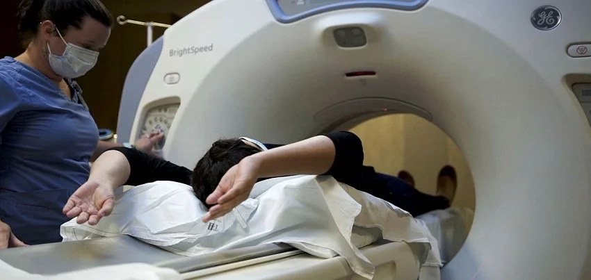
CT scan of the abdomen A CT scan of the abdomen is a medical imaging test that uses X-rays and computer technology to create detailed images of the abdominal area, including organs such as the liver, pancreas, spleen, and kidneys. It is a non-invasive procedure that can help diagnose and monitor a variety of medical conditions. The test is conducted using a specialized machine called a CT scanner. During the procedure, the patient lies on a table that slides into a donut-shaped machine. The scanner rotates around the patient, taking multiple X-ray images from different angles. These images are then processed by a computer to create detailed images of the abdomen. In terms of preparation for the test, there may be specific instructions given by the healthcare provider or imaging facility. For example, patients may be asked to avoid eating or drinking for a certain period of time before the test, or to avoid certain medications. It’s important to follow any instructions provided to ensure the most accurate results. There are a variety of symptoms and conditions that may warrant a CT scan of the abdomen. For example, the test may be ordered to diagnose or monitor a variety of conditions, including abdominal pain, unexplained weight loss, digestive issues, or blood in the stool. The test may also be used to monitor the progression of certain conditions, such as cancer. The time it takes to complete a CT scan of the abdomen can vary depending on the complexity of the imaging needed. In general, the test itself takes only a few minutes, although additional time may be needed for preparation and positioning. Interpreting the results of a CT scan of the abdomen requires specialized training and expertise. The images produced by the test are highly detailed, and it takes a trained healthcare professional to understand what the images show and how they relate to a patient’s specific medical condition. In many cases, the healthcare provider who ordered the test will review the results with the patient and provide an interpretation. In the context of health checkups and wellness programs, CT scans of the abdomen can be a valuable tool for monitoring overall health and identifying potential health issues before they become more serious. Many wellness programs offer CT scans of the abdomen as part of a comprehensive health evaluation, which can help individuals take a proactive approach to their health. Corporate health programs may also incorporate CT scans of the abdomen as a way to monitor the health of employees and identify potential health risks. For example, employers may offer CT scans of the abdomen as part of a preventative health screening program, or as part of a wellness incentive program. By providing employees with access to these types of imaging tests, employers can help employees take a more active role in their health and potentially reduce healthcare costs over the long term. In conclusion, a CT scan of the abdomen is a non-invasive medical imaging test that can help diagnose and monitor a variety of medical conditions. The test is conducted using a specialized machine called a CT scanner, and may require specific preparation depending on the body part being examined. Symptoms that may prompt a healthcare provider to order a CT scan of the abdomen include abdominal pain, unexplained weight loss, digestive issues, or blood in the stool. Interpreting the results of a CT scan of the abdomen requires specialized training and expertise, and the test can be a valuable tool for monitoring overall health and identifying potential health issues before they become more serious.
Read MoreTop rated products
-
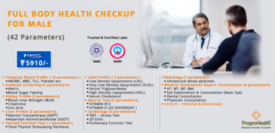
 Full Body Health Checkup I
Original price was: ₹3,000.00.₹1,770.00Current price is: ₹1,770.00.
Full Body Health Checkup I
Original price was: ₹3,000.00.₹1,770.00Current price is: ₹1,770.00.
-
 MFG IND PACK III
Original price was: ₹3,900.00.₹2,145.00Current price is: ₹2,145.00.
MFG IND PACK III
Original price was: ₹3,900.00.₹2,145.00Current price is: ₹2,145.00.
-
 Dengue NS1 Antigen Test
₹900.00
Dengue NS1 Antigen Test
₹900.00
-
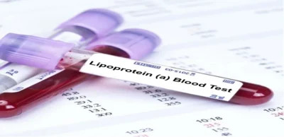 Lipoprotein (A) Blood Test – Lp(a)
₹750.00
Lipoprotein (A) Blood Test – Lp(a)
₹750.00
-
 Healthy Life Advance Male
Original price was: ₹15,000.00.₹8,250.00Current price is: ₹8,250.00.
Healthy Life Advance Male
Original price was: ₹15,000.00.₹8,250.00Current price is: ₹8,250.00.