PrognoHealth – Corporate Health & Wellness Specialist


Ammonia Test Ammonia is a waste product that is produced by the body as a result of the breakdown of proteins. The ammonia test measures the level of ammonia in the blood and is used to assess liver function and to detect certain medical conditions such as liver disease, kidney disease, and metabolic disorders. Pre-test preparation: No special preparation is needed for the ammonia test. Testing method: A blood sample is taken from a patient and sent to the laboratory for analysis. The sample is then analyzed to determine the level of ammonia in the blood. Common symptoms for prescribing this test: The ammonia test is usually ordered when a patient has symptoms of liver or kidney disease such as confusion, disorientation, or mental changes. The test is also used as a follow-up test to monitor treatment of liver or kidney disease, or to detect certain metabolic disorders such as urea cycle disorders. Diagnosis: Elevated ammonia levels can indicate liver or kidney disease, or metabolic disorders, but the diagnosis of these conditions is typically based on a combination of clinical, laboratory, and imaging findings. Reference range: The reference range for ammonia levels can vary depending on the lab, but generally, it is considered normal for adult to have ammonia levels of 15-45 µmol/L. Normal values: The normal range for ammonia levels can vary depending on the lab, but typically falls between 15-45 µmol/L in adults. It is important to note that an elevated ammonia level does not confirm a diagnosis of liver or kidney disease, or metabolic disorders, and should be interpreted along with clinical presentation and other laboratory test results. Additionally, other factors such as age, sex, and certain medications can affect ammonia levels, so the results should be considered in the context of the clinical history and other tests results.
Read More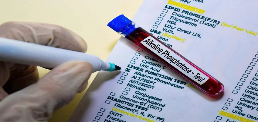
Alkaline Phosphatase (ALP) is an enzyme found in various tissues throughout the body, including the liver, bone, and intestine.
Read More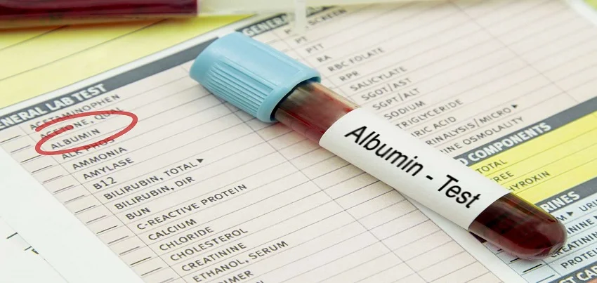
Albumin Test Albumin is a protein made by the liver that plays a critical role in maintaining proper fluid balance in the body. The albumin test measures the level of albumin in the blood and is used to assess liver and kidney function, and to detect malnutrition and other conditions that affect the production or loss of protein in the body. Pre-test preparation: No special preparation is needed for the albumin test. Testing method: A blood sample is taken from a patient and sent to the laboratory for analysis. The sample is then analyzed to determine the level of albumin in the blood. Common symptoms for prescribing this test: The albumin test is usually ordered when a patient has symptoms of liver or kidney disease, malnutrition or protein-losing conditions such as protein-losing enteropathy, nephrotic syndrome, or cirrhosis. The test is also used to monitor the treatment of these conditions. Diagnosis: Low albumin levels can indicate liver or kidney disease, malnutrition or protein-losing conditions, but the diagnosis of these conditions is typically based on a combination of clinical, laboratory, and imaging findings. Reference range: The reference range for albumin levels can vary depending on the lab, but generally, it is considered normal for adult to have albumin levels of 3.5-5.5 g/dL. Normal values: The normal range for albumin levels can vary depending on the lab, but typically falls between 3.5-5.5 g/dL in adults. It is important to note that low albumin levels do not confirm a diagnosis of liver or kidney disease, malnutrition or protein-losing conditions, and should be interpreted along with clinical presentation and other laboratory test results. Additionally, other factors such as age, sex, and certain medications can affect albumin levels, so the results should be considered in the context of the patient’s overall clinical picture. Additionally, Albumin levels can be affected by other factors such as dehydration, infection, surgery, and burns, so it is important to correlate the results with the clinical presentation.
Read More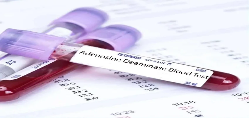
Adenosine Deaminase (ADA) Test Adenosine Deaminase (ADA) is an enzyme that is present in many cells in the body. The ADA test measures the level of ADA in the blood, and it is primarily used to help diagnose certain types of immune deficiencies and to monitor the treatment of those conditions. Pre-test preparation: No special preparation is needed for the ADA test. Testing method: A blood sample is taken from a patient and sent to the laboratory for analysis. The sample is then analyzed to determine the level of ADA in the blood. Common symptoms for prescribing this test: The ADA test is usually ordered when a patient has symptoms of an immune deficiency such as recurrent infections, slow wound healing, and anemia. The test is also ordered as a follow-up test to monitor treatment of an immune deficiency. Diagnosis: Elevated ADA levels can indicate the presence of certain types of immune deficiencies, such as ADA deficiency and SCID (Severe Combined Immune Deficiency). However, the diagnosis of these conditions is typically based on a combination of clinical, laboratory, and genetic tests. Reference range: The reference range for ADA levels can vary depending on the lab, but generally, it is considered normal for adult to have ADA levels of 0-40 U/L. Normal values: The normal range for ADA levels can vary depending on the lab, but typically falls between 0-40 U/L in adults. It is important to note that an elevated ADA level does not confirm a diagnosis of an immune deficiency and should be interpreted along with clinical presentation and other laboratory test results. Additionally, other factors such as age, sex, and certain medications can affect ADA levels, so the results should be considered in the context of the patient’s overall clinical picture. It is also important to note that the ADA test is not a diagnostic test for all types of immune deficiencies and other tests may be needed to confirm the diagnosis. Understanding ADA Test: Normal Range, Purpose, and Adenosine Deaminase Deficiency Introduction: The Adenosine Deaminase (ADA) test is a crucial diagnostic tool used to assess the levels of ADA enzyme in the blood. This test plays a vital role in diagnosing various medical conditions, particularly Adenosine Deaminase Deficiency. In this article, we’ll delve into the significance of the ADA test, its normal range, and its role in identifying ADA deficiency. What is the ADA Test? The ADA test measures the activity of the ADA enzyme in the blood. ADA is an enzyme involved in the breakdown of purines, which are essential components of DNA and RNA. Its primary function is to convert adenosine to inosine and deoxyadenosine to deoxyinosine. This process is crucial for the normal functioning of the immune system, as ADA deficiency can impair lymphocyte development and function. Normal Range of ADA Test: The normal range for ADA activity in the blood varies slightly depending on the laboratory and the method used for testing. However, in general, the normal range for ADA activity in adults is typically between 5 and 40 units per liter (U/L) of blood. Values outside this range may indicate an underlying medical condition, such as ADA deficiency or certain types of cancer. Purpose of ADA Test: The ADA test serves multiple purposes in clinical practice. One of its primary uses is in the diagnosis of ADA deficiency, a rare genetic disorder characterized by low levels of ADA enzyme activity. ADA deficiency can lead to severe combined immunodeficiency (SCID), a life-threatening condition in which the immune system is severely compromised, leaving affected individuals susceptible to recurrent infections. In addition to diagnosing ADA deficiency, the ADA test may also be used to: Monitor the effectiveness of treatment: For individuals undergoing enzyme replacement therapy or bone marrow transplantation for ADA deficiency, regular monitoring of ADA levels can help assess the effectiveness of treatment. Aid in the diagnosis of certain cancers: Elevated ADA levels have been observed in certain types of cancer, such as leukemia and lymphoma. Measuring ADA activity in the blood may therefore help in the diagnosis and monitoring of these conditions. Assist in the diagnosis of tuberculous pleural effusion: ADA levels are often elevated in the pleural fluid of individuals with tuberculous pleural effusion, a form of tuberculosis affecting the lining of the lungs. Measuring ADA activity in pleural fluid can aid in the diagnosis of this condition. Adenosine Deaminase Deficiency: Adenosine Deaminase Deficiency is a rare genetic disorder caused by mutations in the ADA gene, which result in reduced or absent ADA enzyme activity. This deficiency impairs the normal functioning of the immune system, particularly the development and function of T and B lymphocytes. Individuals with ADA deficiency are highly susceptible to recurrent and severe infections, and without treatment, the condition can be fatal in infancy. However, with early diagnosis and appropriate treatment, such as enzyme replacement therapy or bone marrow transplantation, the prognosis for individuals with ADA deficiency has significantly improved. Conclusion: The ADA test is a valuable diagnostic tool with diverse clinical applications, ranging from the diagnosis of ADA deficiency to the monitoring of certain cancers and infectious diseases. By measuring ADA enzyme activity in the blood, healthcare providers can gain valuable insights into the functioning of the immune system and identify underlying medical conditions that may require further evaluation and treatment. Furthermore, for individuals diagnosed with ADA deficiency, early intervention is critical for improving outcomes and preventing complications associated with this rare genetic disorder.
Read More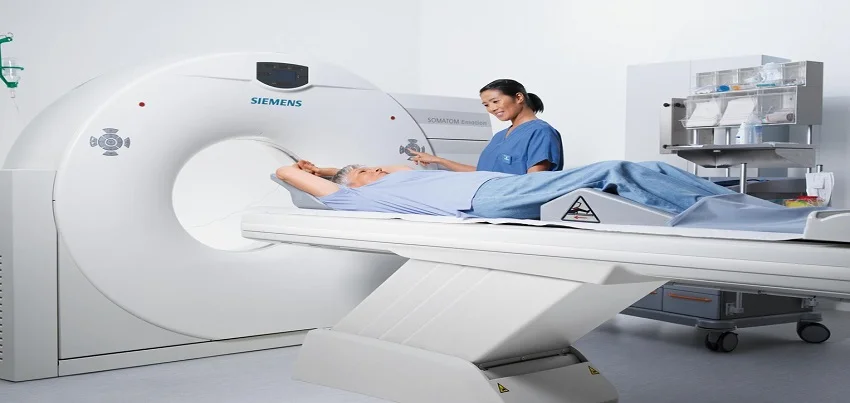
CT Scan Of The Thoracic Spine Test A CT scan of the thoracic spine is a diagnostic imaging test that helps physicians evaluate the bones, discs, and surrounding soft tissues of the middle and upper back. In this blog post, we’ll discuss how the test is conducted, any necessary preparation, common symptoms for ordering the test, and what patients can expect from the results. Firstly, let’s look at how the test is conducted. A CT scan of the thoracic spine typically involves the use of X-rays and computer technology to create detailed, cross-sectional images of the body. During the test, the patient lies on a table that slides into a machine called a CT scanner. The scanner emits X-rays and rotates around the body to capture images from multiple angles. These images are then processed by a computer to create a three-dimensional image of the thoracic spine. As with any medical imaging test, there may be some preparation required. Depending on the reason for the test, patients may be asked to fast for a certain period of time beforehand, avoid taking certain medications, or drink a contrast material that helps to enhance the images. It’s important for patients to follow their doctor’s instructions carefully in order to ensure the most accurate and reliable results. Now, let’s explore the common symptoms that may lead to ordering a CT scan of the thoracic spine. This type of test is often used to diagnose a variety of conditions affecting the middle and upper back, including herniated discs, spinal stenosis, degenerative disc disease, and spinal fractures. Patients experiencing persistent back pain, weakness, numbness, or tingling in the middle or upper back may be candidates for a thoracic spine CT scan. In terms of how long the test takes, a CT scan of the thoracic spine typically takes less than an hour from start to finish. However, patients may be asked to arrive early for preparation or may experience delays depending on the specific facility and equipment being used. Finally, let’s discuss what patients can expect from the results of a thoracic spine CT scan. The images produced during the test can provide a detailed look at the bones, discs, and surrounding soft tissues of the middle and upper back, allowing physicians to identify any abnormalities or issues that may be causing symptoms. Based on the results of the test, doctors may recommend further testing, such as an MRI or a biopsy, or may develop a treatment plan that may include medication, physical therapy, or surgery. In summary, a CT scan of the thoracic spine is a valuable diagnostic tool that can help physicians evaluate a range of conditions affecting the middle and upper back. Patients should follow their doctor’s instructions carefully to prepare for the test and can expect to receive results that provide important information about their health and well-being. As with any medical test, it’s important to discuss any concerns or questions with your doctor in advance.
Read More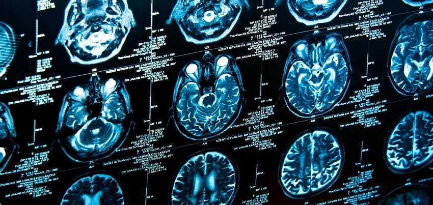
CT Scan Brain A CT scan of the brain is a medical imaging test that is used to create detailed images of the brain. This non-invasive diagnostic test is an important tool that helps doctors diagnose and treat various conditions that affect the brain. In this blog, we will explore what a CT scan of the brain entails, how it is conducted, what symptoms may necessitate the test, and how the results are interpreted. We will also look at how the test fits into the context of health checkups, wellness programs, and corporate health. What is a CT Scan Brain?A CT scan, also known as a computed tomography scan, uses a combination of X-rays and computer technology to produce detailed images of the inside of the body. A CT scan of the brain produces cross-sectional images of the brain that can be used to diagnose a variety of conditions, including tumors, infections, inflammation, bleeding, and injuries.The test works by having the patient lie on a table that slides into a large, circular machine that takes X-ray images of the brain from different angles. The machine then uses these images to create a 3D image of the brain. How is a CT Scan Brain Conducted?The procedure for a CT scan of the brain is simple and non-invasive. A CT scan of the brain is usually performed as an outpatient procedure, meaning that the patient can go home on the same day as the test. Patients will be asked to remove any metal objects, such as jewelry or glasses, and may need to wear a hospital gown during the test.The patient then lies on a table that slides into the CT scanner, which takes a series of X-ray images of the brain. The patient may be asked to hold their breath for a few seconds to ensure the images are clear. The whole procedure usually takes only a few minutes.After the test, the images are sent to a radiologist, who is a specialist in interpreting medical images. The radiologist will analyze the images and write a report of their findings. Test Preparation for a CT Scan Brain:For a CT scan of the brain, there is usually no special preparation required. Patients may be asked to avoid eating or drinking for a few hours before the test, particularly if they are going to receive contrast dye. This dye is injected into a vein in the arm to help enhance the images of the brain.Patients should inform their doctor if they have any allergies, particularly to contrast dye or iodine, as well as if they are pregnant. Common Symptoms for Ordering a CT Scan Brain:A CT scan of the brain may be ordered by a doctor if a patient is experiencing symptoms such as:HeadachesDizzinessSeizuresNausea and vomitingChanges in vision or speechDifficulty moving or weakness in the arms or legsLoss of consciousnessConfusion or memory lossPersonality changes The test can help diagnose conditions such as brain tumors, strokes, bleeding, or swelling in the brain, and infections such as meningitis or encephalitis. Time Taken for the Test and Results Interpretations A CT scan of the brain usually takes only a few minutes. Patients may need to stay still during the scan to ensure clear images. After the test, the images are sent to a radiologist, who will analyze them and write a report of their findings. The results are usually available within a few days. The radiologist will look for any abnormalities in the brain, such as tumors, bleeding, or swelling. They may also look for signs of infections or other conditions that may be affecting the brain.
Read More
Sickling test is a laboratory test used to diagnose sickle cell anemia, a genetic blood disorder characterized by abnormal hemoglobin molecules
Read More
Neonatal Hemoglobin Electrophoresis (Hb Electrophoresis) is a laboratory test
Read More
Blood Sugar Random Test A blood sugar random test measures the amount of glucose (sugar) in a person’s blood at any random time, regardless of when they last ate. There is no specific preparation required for this test, but it is generally recommended to avoid consuming food or drink (other than water) for at least 8 hours before the test. The test is typically performed by taking a small sample of blood from a finger prick or from a vein in the arm. The blood sample is then analyzed using a glucose meter or sent to a laboratory for analysis. The test is commonly prescribed for people who have symptoms of high blood sugar, such as increased thirst, frequent urination, blurred vision, and fatigue. It is also used to diagnose and monitor diabetes, a condition in which the body is unable to properly regulate blood sugar levels. The reference range for blood sugar is typically considered to be between 70 and 100 mg/dL. However, normal values can vary depending on factors such as the person’s age, sex, and overall health. It’s important to note that this test is not a definitive diagnosis and it is recommended to consult with a doctor and get a proper diagnosis with clinical examination and other tests.
Read More
Staying mentally sharp is crucial for success. In today’s fast-paced corporate world age-related memory loss has become a concern.
Read MoreTop rated products
-
 Full Body Health Checkup I
Original price was: ₹3,000.00.₹1,770.00Current price is: ₹1,770.00.
Full Body Health Checkup I
Original price was: ₹3,000.00.₹1,770.00Current price is: ₹1,770.00.
-
 MFG IND PACK III
Original price was: ₹3,900.00.₹2,145.00Current price is: ₹2,145.00.
MFG IND PACK III
Original price was: ₹3,900.00.₹2,145.00Current price is: ₹2,145.00.
-
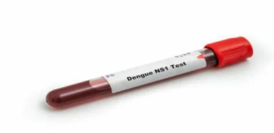 Dengue NS1 Antigen Test
₹900.00
Dengue NS1 Antigen Test
₹900.00
-
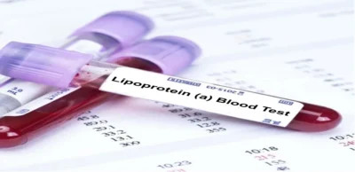 Lipoprotein (A) Blood Test – Lp(a)
₹750.00
Lipoprotein (A) Blood Test – Lp(a)
₹750.00
-
 Healthy Life Advance Male
Original price was: ₹15,000.00.₹8,250.00Current price is: ₹8,250.00.
Healthy Life Advance Male
Original price was: ₹15,000.00.₹8,250.00Current price is: ₹8,250.00.