PrognoHealth – Corporate Health & Wellness Specialist

Amaurosis Fugax Amaurosis fugax is a temporary loss of vision in one eye that is caused by a temporary reduction in blood flow to the eye’s retina. The most common symptoms of amaurosis fugax are a sudden loss of vision in one eye, often accompanied by a visual field defect, such as a dark or blind spot in the field of vision. The vision loss usually lasts for a few minutes to a few hours and typically resolves on its own. To diagnose amaurosis fugax, a healthcare provider will typically perform a thorough eye exam, including visual acuity testing and examination of the eye’s retina. Other tests, such as an ultrasound of the carotid artery, may also be performed to evaluate blood flow to the eye. Treatment for amaurosis fugax typically involves addressing the underlying cause of the reduced blood flow, which can include medications to lower cholesterol or blood pressure, or surgery to repair a narrowed or blocked carotid artery. In some cases, a person with amaurosis fugax may also require antiplatelet therapy or anticoagulation therapy to prevent blood clots. Preventing amaurosis fugax from occurring can be done by controlling blood pressure, cholesterol and glucose level, avoiding smoking and manage other health conditions that may affect blood flow such as diabetes and atherosclerosis. Corporate health & wellness programs can play a role in preventing amaurosis fugax by educating employees on the importance of maintaining healthy lifestyle choices, such as regular exercise, healthy diet, and regular check-ups, to manage blood pressure, cholesterol and glucose level, and also providing access to resources for health screenings and preventive care. In summary, Amaurosis Fugax is a temporary loss of vision in one eye caused by a temporary reduction in blood flow to the eye’s retina. The most common symptoms of Amaurosis Fugax are a sudden loss of vision in one eye, often accompanied by a visual field defect. To diagnose Amaurosis Fugax, a healthcare provider will typically perform a thorough eye exam and other tests such as an ultrasound of the carotid artery. Preventing Amaurosis Fugax from occurring can be done by controlling blood pressure, cholesterol and glucose level, avoiding smoking and manage other health conditions that may affect blood flow such as diabetes and atherosclerosis. Corporate health & wellness programs can play a role in preventing amaurosis fugax by educating employees on the importance of maintaining healthy lifestyle choices, such as regular exercise, healthy diet, and regular check-ups, to manage blood pressure, cholesterol and glucose level, and also providing access to resources for health screenings and preventive care.
Read More
Creatinine Test The Creatinine test measures the level of creatinine in the blood. Creatinine is a waste product that is produced by muscle metabolism, and it is normally removed from the body by the kidneys. The creatinine test is used to evaluate kidney function and to detect and monitor kidney disease. Pre-test preparation: There is no specific preparation required for a Creatinine test. Testing method: The Creatinine test is a blood test. A healthcare provider will take a sample of blood from the patient’s arm, usually from a vein in the elbow or the back of the hand. The blood sample will be sent to a laboratory for analysis. Symptoms for prescribing this test: The Creatinine test may be ordered if a person has symptoms of kidney disease such as fatigue, weakness, decreased urine output, or swelling in the legs and ankles, or if a healthcare provider suspects an underlying condition that may affect kidney function. Diagnosis: Creatinine test results can be used, along with other tests such as a urinalysis and a creatinine clearance test, to diagnose and monitor kidney disease, and to evaluate the effectiveness of treatment. Reference range and normal values: The normal range for creatinine levels in the blood varies depending on the laboratory that performs the test, but it typically ranges between 0.5-1.3 mg/dL for men and 0.4-1.2 mg/dL for women. Medical disclaimer: It is important to note that a creatinine test is just one aspect of a diagnosis and that other tests and factors will be considered. It is also important to consult your healthcare provider for professional and personalized advice. The information provided is not intended to be a substitute for professional medical advice, diagnosis, or treatment. Always seek the advice of your physician or other qualified healthcare provider with any questions you may have regarding a medical condition
Read More
C-Peptide Test The C-Peptide test measures the level of C-peptide in the blood. C-peptide is a byproduct of insulin production, and it is secreted into the bloodstream in equal amounts to insulin. The C-peptide test is used to help diagnose and monitor diabetes, specifically to differentiate between type 1 and type 2 diabetes and to monitor the effectiveness of insulin treatment in type 1 diabetes. Pre-test preparation: There is no specific preparation required for a C-Peptide test. Testing method: The C-Peptide test is a blood test. A healthcare provider will take a sample of blood from the patient’s arm, usually from a vein in the elbow or the back of the hand. The blood sample will be sent to a laboratory for analysis. Symptoms for prescribing this test: The C-Peptide test may be ordered if a person has symptoms of diabetes such as frequent urination, excessive thirst, and weight loss, or if a healthcare provider suspects an underlying condition that may affect insulin production. Diagnosis: C-Peptide test results can be used, along with other tests such as glucose and HbA1c, to diagnose and monitor diabetes, and to evaluate the effectiveness of insulin treatment in type 1 diabetes. Reference range and normal values: The normal range for C-Peptide levels in the blood is typically between 0.5-2.5 ng/mL. However, this may vary slightly depending on the laboratory that performs the test. Medical disclaimer: It is important to note that a C-Peptide test is just one aspect of a diagnosis and that other tests and factors will be considered. It is also important to consult your healthcare provider for professional and personalized advice. The information provided is not intended to be a substitute for professional medical advice, diagnosis, or treatment. Always seek the advice of your physician or other qualified healthcare provider with any questions you may have regarding a medical condition
Read More
Silicosis Silicosis is a lung disease caused by inhaling dust containing silica particles. The dust causes inflammation and scarring in the lungs, which can make it difficult to breathe. Silicosis is a serious condition that can lead to disability and even death. Symptoms of silicosis include shortness of breath, chest tightness, and a persistent cough. As the disease progresses, the affected person may also experience fatigue, weight loss, and an overall decline in health. In advanced cases, the person may develop lung infections, such as tuberculosis. Diagnosis of silicosis involves a physical exam, chest X-ray, and CT scan. A lung function test may also be done to determine how well the lungs are working. In some cases, a biopsy of the lung tissue may be needed to confirm the diagnosis. Common treatment methods for silicosis include medications to reduce inflammation and help open up the airways. Oxygen therapy may also be used to help the person breathe easier. In advanced cases, surgery may be needed to remove damaged lung tissue. Preventing silicosis from occurring is key to protecting the health of workers and others who may be exposed to silica dust. This can be done by using proper ventilation and personal protective equipment, such as respirators, when working with silica dust. Employers should also make sure that workers are properly trained on how to handle silica dust safely. Annual health check-ups and corporate health & wellness programs can play a crucial role in preventing silicosis. These check-ups can help identify potential health risks, such as exposure to silica dust, and provide workers with the resources and support they need to stay healthy. Corporate health & wellness programs can also help promote healthy habits, such as regular exercise and a healthy diet, which can help reduce the risk of silicosis and other lung diseases. In terms of diet and exercise, a healthy diet with plenty of fruits and vegetables can help improve lung function and strengthen the immune system. Regular exercise, such as brisk walking or cycling, can also help improve lung function and reduce the risk of lung diseases. In conclusion, Silicosis is a serious lung disease caused by inhaling silica dust. It can cause inflammation and scarring in the lungs, making it difficult to breathe. Symptoms include shortness of breath, chest tightness, and a persistent cough. To prevent silicosis from occurring, proper ventilation and personal protective equipment, such as respirators, should be used when working with silica dust. Annual health check-ups and corporate health & wellness programs can play a crucial role in preventing silicosis and promoting overall health. A healthy diet and regular exercise can also help reduce the risk of silicosis and other lung diseases.
Read More
Haemophilia Haemophilia is a rare genetic disorder that affects the blood’s ability to clot properly. It is caused by a deficiency or malfunction of certain clotting factors in the blood, which leads to excessive bleeding and bruising. There are two main types of haemophilia: haemophilia A (also known as classic haemophilia), which is caused by a deficiency of clotting factor VIII, and haemophilia B (also known as Christmas disease), which is caused by a deficiency of clotting factor IX. Symptoms of haemophilia can vary depending on the severity of the condition, but may include excessive bleeding and bruising, prolonged bleeding from cuts or injuries, joint pain and stiffness, and nosebleeds. In severe cases, haemophilia can lead to life-threatening bleeding in the brain, joints, or other vital organs. Diagnosis of haemophilia is typically made through a blood test to measure the levels of clotting factors in the blood. Genetic testing may also be used to confirm the diagnosis and identify the specific type of haemophilia. Common treatment methods for haemophilia include replacement therapy, which involves administering the missing clotting factor to the patient through regular injections or infusions. In some cases, surgery may be necessary to control bleeding or repair damaged joints. Preventing haemophilia is not possible as it is a genetic disorder, but early diagnosis and appropriate treatment can help to prevent serious complications and improve quality of life. During an annual health check-up, it is important for individuals with a family history of haemophilia to be screened for the condition. Corporate health and wellness programs can also play a role in supporting employees who have haemophilia by providing access to appropriate care and resources. Although, diet and exercise do not prevent haemophilia, but it can help to maintain overall health and well-being. A healthy diet that is high in fruits, vegetables, and lean proteins can help to support the body and improve overall health. Exercise can also help to maintain joint mobility, improve cardiovascular health, and reduce stress. In conclusion, Haemophilia is a rare genetic disorder that affects the blood’s ability to clot properly. By understanding the symptoms, getting regular check-ups and appropriate treatment, it is possible to prevent serious complications and improve quality of life. Corporate health and wellness programs can also play a key role in supporting employees who have haemophilia by providing access to appropriate care and resources.
Read More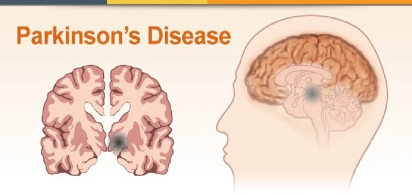
Parkinson’s Disease (PD) Parkinson’s disease (PD) is a progressive disorder of the nervous system that affects movement. It is characterised by tremors, stiffness, and difficulty with coordination and balance. The disease is caused by the degeneration of dopamine-producing cells in the brain. Symptoms of PD can vary from person to person and can include tremors, stiffness, difficulty with coordination and balance, and a slow or shuffling gait. Other symptoms may include difficulty with fine motor skills, such as buttoning clothes or writing, and changes in speech and voice. Some people with PD may also experience depression, anxiety, and cognitive changes. To diagnose PD, a healthcare provider will typically perform a physical examination, review the patient’s medical history, and conduct a series of neurological tests. These tests may include a movement disorder examination, a test of reflexes, and an examination of the patient’s balance and coordination. In some cases, imaging tests such as an MRI or a CT scan may be performed. Common treatment methods for PD include medication, physical therapy, and surgery. Medications, such as levodopa and dopamine agonists, can help to increase the levels of dopamine in the brain and improve symptoms. Physical therapy can help to improve balance and coordination, and may also help to reduce stiffness and tremors. Surgery, such as deep brain stimulation, may be used in some cases to improve symptoms that do not respond to other treatments. To prevent PD from occurring, it is important to maintain a healthy lifestyle. This includes eating a healthy diet and getting regular exercise. Annual health check-ups are also important to detect and prevent any abnormal changes or conditions. Corporate health and wellness programs can also help to educate and encourage employees to maintain a healthy lifestyle. Diet and exercise can play a significant role in preventing PD. Eating a diet that is rich in fruits, vegetables, and whole grains can help to reduce the risk of developing PD. In addition, regular exercise can help to maintain a healthy weight and reduce the risk of developing certain health conditions. Research suggests that maintaining a healthy lifestyle, eating a well-balanced diet, regular exercise and avoiding environmental toxins may reduce the risk of developing PD. There is some evidence that suggests that smoking, exposure to pesticides, and head injuries may increase the risk of developing PD. In conclusion, Parkinson’s Disease is a progressive disorder of the nervous system that affects movement. Symptoms vary from person to person and can include tremors, stiffness, difficulty with coordination and balance, and a slow or shuffling gait. PD is diagnosed through physical examination, medical history review and neurological tests. Common treatment methods include medication, physical therapy and surgery. To prevent PD from occurring, it is important to maintain a healthy lifestyle, eating a well-balanced diet, regular exercise and avoiding environmental toxins. Corporate health and wellness programs can also help to educate and encourage employees to maintain a healthy lifestyle. Regular check-ups and screenings are also important in order to detect any abnormal changes or conditions early on.
Read More
Dental CT Scan of the Mandible. A dental CT scan of the mandible, also known as a dental cone beam computed tomography (CBCT) scan, is a specialised imaging technique that produces detailed images of the mandible or lower jaw. The test is commonly used to diagnose and evaluate dental conditions, such as impacted teeth, jaw tumors, and temporomandibular joint (TMJ) disorders. In this article, we will discuss the procedure of the Dental CT Scan Mandible, test preparation, common symptoms for ordering the test, time taken for the test, result interpretation, and its importance in corporate health wellness packages. Test Procedure: During a Dental CT Scan Mandible, the patient sits or lies down on a dental chair, and a specialized CBCT scanner is placed around the patient’s head to take multiple images of the mandible. The scanner rotates around the head, taking images from different angles to create a three-dimensional image of the mandible. The entire procedure usually takes between 10 to 20 minutes. Test Preparation: Patients do not require any special preparation for a Dental CT Scan Mandible. They should wear comfortable clothing and remove any metal objects, such as jewelry or hairpins, as these can interfere with the imaging process. Patients should inform their doctor of any allergies they have or any previous surgeries or medical procedures they have undergone. A Dental CT Scan Mandible may be ordered by a dentist or oral surgeon to investigate the following symptoms or conditions:Impacted teethJaw tumorsTMJ disordersTooth infections or abscessesTooth loss or damageEvaluation for dental implants or orthodontic treatment Time Taken for the Test and its Results Interpretation: The Dental CT Scan Mandible usually takes between 10 to 20 minutes to complete. After the test, a radiologist or oral radiologist will interpret the images and provide a report to the referring dentist or oral surgeon. The report will include information about the condition of the mandible, including any abnormalities such as tumors, cysts, or fractures. Importance in Corporate: Health Wellness Packages Dental CT Scan Mandible is a critical diagnostic tool that can detect and diagnose dental conditions that may not be visible through traditional dental X-rays. Early detection and treatment of these conditions can help prevent complications, reduce the need for more invasive procedures, and ultimately lead to better dental health outcomes. By offering Dental CT Scan Mandible as part of corporate health wellness packages, companies can help their employees detect and diagnose these conditions early, leading to better overall health and productivity. In conclusion, Dental CT Scan Mandible is a specialized imaging technique used to diagnose and evaluate dental conditions of the mandible. Patients do not require any special preparation for this test, but they should inform their doctor of any allergies they have or any previous surgeries or medical procedures they have undergone. Results of the test are interpreted by a radiologist or oral radiologist and provided to the referring dentist or oral surgeon. Companies can offer Dental CT Scan Mandible as part of their corporate wellness packages to help their employees detect and diagnose these conditions early and promote better overall dental health and productivity.
Read More
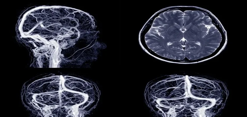
MRI of the Lumbar and Sacral Spine Magnetic Resonance Imaging (MRI) of the Lumbar and Sacral Spine is a non-invasive diagnostic test that uses a powerful magnetic field, radio waves, and a computer to produce detailed images of the lower back and pelvis. The test is conducted to evaluate a range of conditions affecting the lumbar and sacral spine, including injuries, disc herniation, spinal stenosis, tumors, and other abnormalities. Test Conducted: During an MRI of the Lumbar and Sacral Spine, the patient lies on a table that slides into a cylindrical machine that houses the MRI scanner. The scanner uses a magnetic field and radio waves to create images of the lumbar and sacral spine. The test typically takes between 30 and 60 minutes to complete, depending on the complexity of the exam and the patient’s ability to remain still during the procedure. Test Preparation: For an MRI of the Lumbar and Sacral Spine, patients should wear comfortable, loose-fitting clothing without metal zippers or buttons. The patient will be asked to remove any metal objects, such as jewelry, watches, or belts. In some cases, the patient may need to fast for a few hours before the exam, depending on the reason for the test. Common Symptoms for Ordering the Test: MRI of the Lumbar and Sacral Spine may be ordered by a physician if a patient has symptoms such as lower back pain, leg pain, numbness or tingling in the legs or feet, weakness in the legs or feet, or difficulty standing or walking. The test can help diagnose a range of conditions affecting the lumbar and sacral spine, including injuries, disc herniation, spinal stenosis, tumors, and other abnormalities. Time Taken for the Test and Interpretation of Results: An MRI of the Lumbar and Sacral Spine typically takes between 30 and 60 minutes to complete, and patients can return to their normal activities immediately after the test. The images produced by the MRI are examined by a radiologist, who will interpret the results and provide a report to the ordering physician. The physician will then review the results with the patient and determine the appropriate course of treatment based on the findings.MRI of the Lumbar and Sacral Spine is an important tool for diagnosing and treating a range of conditions affecting the lower back and pelvis, and can be used in regular health checkups and corporate wellness programs. Early detection of lumbar and sacral spine problems is critical to maintaining good health and wellness. In addition, some corporate health programs offer wellness screenings that include lower back exams, and an MRI of the Lumbar and Sacral Spine may be ordered as part of a comprehensive wellness evaluation. In conclusion, MRI of the Lumbar and Sacral Spine is a valuable diagnostic tool that can help detect a range of lower back and pelvis conditions that can affect overall health and wellness. The test is non-invasive and typically takes between 30 and 60 minutes to complete, with some preparation required such as removing metal objects and fasting for a few hours before the exam. The results are interpreted by a radiologist and reviewed by the ordering physician, who will determine the appropriate course of treatment based on the findings. Regular health checkups and corporate wellness programs can also incorporate lower back exams, including MRI of the Lumbar and Sacral Spine, to help detect and manage lumbar and sacral spine problems.
Read More
Lid Imbrication Syndrome Lid Imbrication Syndrome, also known as eyelid malposition, is a condition in which the eyelids are not properly aligned, resulting in eyelid overlap or eyelid retraction. This can lead to a variety of symptoms, including dry eyes, irritation, and even vision loss. Symptoms of Lid Imbrication Syndrome can vary depending on the severity of the condition. They can include dry eyes, itching, redness, blurred vision, and even double vision. In severe cases, the eyelids can become so misaligned that they can rub against the cornea, leading to corneal damage. Diagnosis of Lid Imbrication Syndrome is typically made through a physical examination, during which the physician will look for signs of eyelid misalignment and assess the affected area for any changes in skin texture or color. They may also use a special instrument called a Hertel exophthalmometer to measure the distance between the cornea and the eyelid. Other diagnostic tests that may be used include eyelid photography, which uses a special camera to capture images of the eyelids, and corneal topography, which uses a special device to map the shape of the cornea. Treatment for Lid Imbrication Syndrome can vary depending on the underlying cause of the condition, but common treatment methods include eyelid surgery, also known as blepharoplasty. This procedure involves repositioning the eyelids to their proper position. In some cases, additional procedures, such as Botox injections, or temporary eyelid taping, may be used to help the eyelids heal. Prevention of Lid Imbrication Syndrome is the key to avoid the condition and its symptoms. This is where annual health check-ups and corporate health & wellness programs come in. These programs can help you identify any potential risks for Lid Imbrication Syndrome, such as a family history of the condition, and take steps to prevent it from occurring. For example, if you are at risk for Lid Imbrication Syndrome, your doctor may recommend that you avoid certain activities that could put extra pressure on your eyelids, such as heavy lifting or carrying heavy bags. In addition to taking care of your health, there are also a few lifestyle changes that can help to prevent Lid Imbrication Syndrome. One of the most important things you can do is to maintain a healthy diet and exercise regularly. Eating a diet that is low in fat and high in fiber can help to reduce your risk of Lid Imbrication Syndrome, as can regular exercise, which can help to improve the function of your eyelids. In conclusion, Lid Imbrication Syndrome is a condition in which the eyelids are not properly aligned, resulting in eyelid overlap or eyelid retraction. It can cause a variety of symptoms, including dry eyes, irritation, and even vision loss. It can be diagnosed through physical examination and other diagnostic tests. Treatment options include eyelid surgery, Botox injections, or temporary eyelid taping. To prevent Lid Imbrication Syndrome, it is essential to take care of your health through annual health check-ups and corporate health & wellness programs, maintain a healthy diet and exercise regularly.
Read More
Leukemia: Types, Symptoms, Diagnosis & Treatment Leukemia is a type of cancer that affects the blood and bone marrow. It is characterized by the overproduction of abnormal white blood cells, which can crowd out normal cells and make it difficult for the body to fight infection. There are several types of leukemia, including acute lymphocytic leukemia (ALL), acute myeloid leukemia (AML), chronic lymphocytic leukemia (CLL), and chronic myeloid leukemia (CML). The symptoms and treatment options can vary depending on the type of leukemia. Symptoms of leukemia can include fatigue, weakness, weight loss, easy bruising or bleeding, frequent infections, and swollen lymph nodes. Some types of leukemia may also cause pain in the bones or joints. Diagnosis of leukemia is typically made based on the patient’s symptoms, a physical examination, and laboratory tests such as a complete blood count (CBC) and a bone marrow biopsy. Common treatment methods for leukemia include chemotherapy, radiation therapy, and bone marrow transplant. In some cases, targeted therapy and immunotherapy are also used. The type of treatment will depend on the type of leukemia and the stage of the disease. It is important to note that the cause of leukemia is not fully understood and it’s not known how to prevent it from occurring. However, there are some risk factors that have been identified such as exposure to certain chemicals, radiation exposure, and certain genetic conditions. In terms of diet and exercise, it is essential to eat a balanced diet that is high in fruits and vegetables, and low in processed foods and sugar. Regular exercise, such as going for a daily walk or jog, can help to boost the immune system and reduce stress, which can be beneficial for people with leukemia. Annual health check-ups are also important for detecting any signs of leukemia early on. Corporate health and wellness programs can also be implemented to promote healthy lifestyles and to increase awareness about the disease. It is important to note that there is currently no known way to prevent leukemia. However, it’s important to be aware of the possible risk factors and take precautions to minimize the risk. This can include reducing exposure to chemicals and radiation, and getting regular health check-ups. In addition to the above-mentioned preventive measures, it’s also important to have regular check-ups with a healthcare provider in order to detect any signs of leukemia early on. Corporate health and wellness programs can also play a major role in promoting healthy lifestyles among employees and providing support and resources for individuals who have been diagnosed with leukemia. Overall, leukemia is a type of cancer that affects the blood and bone marrow. There is currently no known way to prevent it, but by being aware of the possible risk factors, practicing healthy lifestyle habits, and having regular check-ups, the risk of developing leukemia can be reduced. Corporate health and wellness programs can also play a major role in promoting healthy lifestyles and increasing awareness about the disease. Leukemia is a complex and often misunderstood disease, categorized as a type of cancer that affects the blood and bone marrow. This condition is characterized by the uncontrolled proliferation of abnormal white blood cells, which impairs the body’s ability to fight infections and hinders the normal function of blood cells. Let’s delve into the various aspects of leukemia, exploring its types, causes, symptoms, and treatments. Types of Leukemia Leukemia is broadly classified into four main types: 1.Acute Lymphocytic Leukemia (ALL): This type is most common in children but can also affect adults. It progresses rapidly and requires prompt treatment.2. Acute Myeloid Leukemia (AML): AML is more common in adults and progresses quickly, necessitating immediate medical intervention.3. Chronic Lymphocytic Leukemia (CLL): This form of leukemia typically affects older adults and progresses slowly, often being diagnosed during routine check-ups before symptoms appear.4. Chronic Myeloid Leukemia (CML): CML is also more prevalent in adults and progresses more slowly than acute forms of leukemia. Causes of Leukemia The exact cause of leukemia is not entirely understood, but several factors may contribute to its development: • Genetic Predisposition: A family history of leukemia can increase the risk.• Exposure to Radiation and Chemicals: High levels of radiation or exposure to certain chemicals like benzene have been linked to leukemia.• Previous Cancer Treatment: Individuals who have undergone chemotherapy or radiation therapy for other cancers have an increased risk.• Genetic Disorders : Conditions such as Down syndrome are associated with a higher incidence of leukemia. Symptoms of Leukemia Leukemia symptoms can vary depending on the type and stage of the disease. However, common symptoms include: • Fatigue and Weakness: Persistent tiredness not alleviated by rest.• Frequent Infections : Due to a compromised immune system.• Fever and Chills: Often without an apparent infection.• Easy Bleeding or Bruising: Minor injuries can result in significant bruising or bleeding.• Swollen Lymph Nodes: Particularly in the neck, underarm, or groin.• Bone or Joint Pain: Due to the spread of leukemia cells in these areas.• Weight Loss: Unintentional and significant weight loss. Early Signs of Leukemia Recognizing the early signs of leukemia can be crucial for timely diagnosis and treatment. These early signs might include: • Frequent Nosebleeds• Night Sweats• Paleness• Shortness of Breath• Petechiae: Small red spots on the skin caused by bleeding. Lymphocytic Leukemia Symptoms Lymphocytic leukemia, whether chronic or acute, often presents with specific symptoms such as: • Swelling of the Liver or Spleen: Leading to abdominal discomfort.• Enlarged Lymph Nodes: Especially in the neck and underarms.• Anemia: Leading to pallor and fatigue. Diagnosis and Leukemia Therapy Diagnosing leukemia typically involves a combination of physical examinations, blood tests, and bone marrow biopsies. Once diagnosed, the treatment plan is tailored based on the type and stage of leukemia. • Chemotherapy: The primary treatment for most types of leukemia, involving drugs that kill or stop the growth of cancer cells.• Radiation Therapy: Uses high-energy radiation to target and destroy leukemia cells.• Stem Cell Transplant: Replaces diseased bone marrow with healthy stem cells to restore normal blood cell production.•
Read MoreTop rated products
-
 Full Body Health Checkup I
Original price was: ₹3,000.00.₹1,770.00Current price is: ₹1,770.00.
Full Body Health Checkup I
Original price was: ₹3,000.00.₹1,770.00Current price is: ₹1,770.00.
-
 MFG IND PACK III
Original price was: ₹3,900.00.₹2,145.00Current price is: ₹2,145.00.
MFG IND PACK III
Original price was: ₹3,900.00.₹2,145.00Current price is: ₹2,145.00.
-
 Dengue NS1 Antigen Test
₹900.00
Dengue NS1 Antigen Test
₹900.00
-
 Lipoprotein (A) Blood Test – Lp(a)
₹750.00
Lipoprotein (A) Blood Test – Lp(a)
₹750.00
-
 Healthy Life Advance Male
Original price was: ₹15,000.00.₹8,250.00Current price is: ₹8,250.00.
Healthy Life Advance Male
Original price was: ₹15,000.00.₹8,250.00Current price is: ₹8,250.00.